Dental bonding is one of the most widely used procedures in restorative and cosmetic dentistry and one of the most misunderstood in terms of what actually makes it work. The term covers two distinct but related applications: direct composite bonding, where resin is applied and shaped directly on the tooth; and adhesive cementation, where a resin-based cement bonds a laboratory-fabricated restoration to tooth structure. Both rely on the same fundamental chemistry, but the protocols, materials, and clinical outcomes differ significantly.
This guide covers dental bonding from both a clinical and a dental lab perspective what it is, how the bonding chemistry works, where it's used, which resin types are involved, and how bonding protocols connect to the ceramic and zirconia restorations that dental labs fabricate. It's written for dental professionals who want technical clarity, not a patient brochure.
What dental bonding actually is?
At its core, dental bonding is the adhesion of a resin-based material to tooth structure enamel, dentine, or both through a combination of micromechanical retention and chemical adhesion. The word "bonding" in dentistry refers specifically to this resin-mediated adhesion mechanism, which distinguishes it from the mechanical retention that older cementation techniques (zinc phosphate, glass ionomer without adhesive) relied on.
The clinical significance of adhesive bonding is substantial. A restoration that bonds chemically and micromechanically to tooth structure distributes load differently than one held by friction and compressive forces alone. For ceramic restorations particularly thin, conservative preparations like veneers and inlays adhesive bonding is what provides the mechanical support that makes the restoration clinically viable. A lithium disilicate veneer at 0.3 mm thickness would fracture under normal function without the reinforcing effect of the adhesive bond to the underlying enamel.
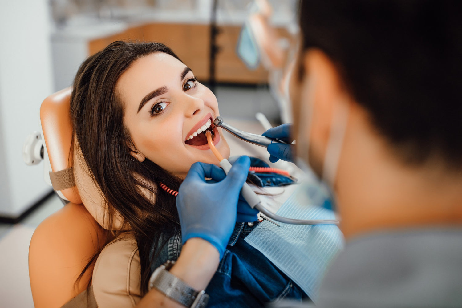
Direct composite bonding: the clinical procedure
Direct dental bonding resin the procedure most patients associate with "bonding" — involves applying composite resin directly to the tooth surface, shaping it, and curing it with a visible-light curing unit. It's used for chipped or fractured teeth, closing diastemas, masking discolouration, and reshaping minor morphological irregularities without tooth preparation.
The procedure follows a consistent protocol:
Etching.
The tooth surface is conditioned with phosphoric acid (typically 35–37%) for 15–30 seconds on enamel and 10–15 seconds on dentine. Acid etching creates a microporous surface in enamel by selectively dissolving hydroxyapatite the roughened surface provides the micromechanical retention that the resin infiltrates and locks into after curing. On dentine, etching is more technique-sensitive because it removes the smear layer and opens dentinal tubule the roughened surface provides the micromechanical retention that the resin infiltrates and locks into after curing. On dentine, etching is more technique-sensitive because it removes the smear layer and opens dentinal tubules, which requires the surface to remain moist for optimal resin infiltration with wet-bonding systems.
Bonding agent application.
A bonding agent a low-viscosity resin primer that wets the etched surface and penetrates the micro-porosity is applied and light-cured before the composite resin is placed. The bonding agent creates the hybrid layer: a zone of co-mingled resin and demineralised collagen at the dentine surface that forms the mechanical foundation of the adhesive joint.
Composite placement and curing.
The composite resin is applied in increments, shaped to the desired morphology, and cured in layers to minimise polymerisation shrinkage stress. Each increment is typically 2mm or less to ensure adequate light penetration and complete polymerisation through the full depth of the resin.
Finishing and polishing.
The cured composite is refined with finishing burs and polished to final surface texture. Surface finish has a direct effect on the restoration's stain resistance and longevity a smooth, well-polished surface accumulates significantly less extrinsic staining than a rough one.
Adhesive cementation: bonding laboratory restorations
Adhesive cementation is the dental lab-relevant application of dental bonding the protocol used to bond ceramic, composite, or other indirect restorations fabricated in the lab to prepared tooth structure. This is where bonding resin selection and protocol compliance have the most direct impact on restoration longevity.
The cementation protocol depends on the restoration material, and this is where the distinction between ceramic types matters practically.
Bonding to lithium disilicate and glass ceramics
Lithium disilicate the material used for anterior veneers, inlays, and single-unit crowns is bondable through both micromechanical and chemical mechanisms. The glass phase of the material is etchd with hydrofluoric acid (5% HF, 20 seconds for IPS e.max CAD; 60 seconds for pressed), which creates a microporous surface similar to acid-etched enamel. Silanation follows — a silane coupling agent creates a chemical bridge between the ceramic surface and the resin cement — and then resin cement is applied and cured.
The combination of micromechanical retention from HF etching and chemical bonding from silane produces bond strengths that meaningfully reinforce the restoration against fracture under occlusal load.This is why cementation protocol compliance is inseparable from lithium disilicate restoration a crown placed without HF etching and silanation loses most of this reinforcing effect and is mechanically compromised at delivery regardless of lab fabrication quality.
why it's different?
Zirconia dental material cannot be etched with hydrofluoric acid the polycrystalline ceramic structure doesn't have a glass phase to dissolve. This means the HF etching nd silanation protocol used for lithium disilicate doesn't work for zirconia, and a different bonding strategy is required.
Current evidence supports two approaches for bonding to zirconia: sandblasting with alumina particles (50 µm Al₂O₃ at 2.5 bar) to create micromechanical retention, followed by application of an MDP-containing primer (10-methacryloyloxydecyl dihydrogen phosphate) that forms a chemical bond to the zirconium oxide surface; or use of a self-adhesive resin cement containing MDP, which combines the cementation and priming steps.
For dental labs fabricating zirconia dental restorations, it's worth including sandblasting instructions and recommended primer products in the case documentation for every zirconia crown or bridge clinicians who aren't familiar with zirconia-specific bonding protocols sometimes apply the lithium disilicate protocol incorrectly, which produces inadequate bond strength.
The zirconia multilayer anterior restorations that most digital labs now fabricate producing translucency levels that approach lithium disilicate still require the zirconia-specific MDP bonding protocol, not HF etching. The optical properties have changed with multilayer formulations; the surface chemistry has not.
Types of dental bonding resin systems
Bonding agents are classified by generation and by etching strategy. For clinical and lab professionals, the most relevant practical distinction is between three main strategies currently in use:
Etch-and-rinse systems (also called total-etch or three-step) apply phosphoric acid separately, rinse it off, then apply primer and adhesive in sequential steps. They produce reliable bond strengths to enamel and dentine but are technique-sensitive particularly the wet-bonding requirement on etched dentine. These systems have the longest clinical track reco particularly the wet-bonding requirement on etched dentine. These systems have the longest clinical track record and the strongest evidence base for enamel bonding.
Self-etch systems combine the etching and priming steps, using acidic monomers that simultaneously condition the surface and infiltrate it without a separate rinse step. They are less technique-sensitive than total-etch on dentine, produce a milder etch that preserves more collagen structure, and are faster to apply. Bond strengths to enamel are generally slightly lower than with phosphoric acid etching a a clinically significant consideration for enamel-dominant preparations.
Universal adhesives can be used in total-etch, selective-etch, or self-etch mode depending on the clinical situation and the clinician's preference. Their MDP content makes them compatible with zirconia bonding when used with appropriate surface preparation. They are the most versatile option in contemporary restorative workflows.
For resin cements specifically the luting agents used to seat laboratory restorations options include dual-cure resin cements (light-cured through the restoration and self-cured where light can't reach), self-adhesive resin cements (no separate adhesive required, suitable for zirconia and metal), and conventional resin cements (require a separate bonding agent).
Clinical indications: where dental bonding is used
Direct composite bonding — minor cosmetic corrections to tooth shape, size, and colour; repair of chipped or fractured anterior teeth; closing small diastemas; masking mild discolouration not amenable to bleaching. Best suited to cases where the extent of change required is modest and the patient's occlusion is favourable. Not appropriate for extensive reshaping, severely discoloured teeth, or patients with parafunctional habits where the composite's lower hardness compared to ceramic becomes a durability concern.
Adhesive cementation of ceramic veneers — lithium disilicate or feldspathic porcelain veneers bonded to minimally prepared or unprepared enamel. The adhesive bond to enamel is the primary mechanical support for the restoration. Preservation of enamel at the preparation margin is therefore a clinical requirement for long-term veneer success bonding to dentine produces lower bond strengths and greater sensitivity risk.
Adhesive cementation of ceramic inlays and onlays — conservative posterior restorations where remaining tooth structure is reinforced by the adhesive bond rather than weakened by crown preparation. Bond strength in this application replaces the retentive geometry that conventional cementation relied on.
Cementation of zirconia crowns and bridges — using MDP-based resin cements or self-adhesive cements after appropriate surface preparation. Posterior zirconia crowns in conventional preparations can also be placed with high-strength conventional cements where adhesive bonding isn't the primary retention mechanism but for anterior positions and single implant crowns, resin cement with MDP primer is the recommended approach.
Why bonding protocol matters for dental labs?
From a dental lab perspective, the bonding protocol used by the clinician directly affects the clinical performance of every restoration the lab fabricates. A lithium disilicate crown that fractures six months post-cementation isn't necessarily a lab fabrication failure it may be an etching and silanation failure at the chair. A zirconia crown that debonds repeatedly may reflect incorrect cement selection rather than marginal fit issues it may be an etching and silanation failure at the chair. A zirconia crown that debonds repeatedly may reflect incorrect cement selection rather than marginal fit issues.
Labs that include material-specific cementation instructions with every case — recommending the correct etching protocol for lithium disilicate, the correct MDP primer protocol for zirconia, and the appropriate cement type — reduce clinician errors that would otherwise result in remakes attributed to the lab. This communication is part of delivering a complete restoration, not an optional extra.
As a dental lab material supplier serving North American laboratories, Zirconia Guys supplies both the Aidite zirconia range covering dental zirconia discs, multilayer, pre-shaded, and white options and related dental lab materials for complete digital workflows. Labs that want to discuss zirconia material selection, zirconia blocks price across the range, or how different zirconium dental material grades interact with cementation protocols are welcome to get in touch with the team directly.


